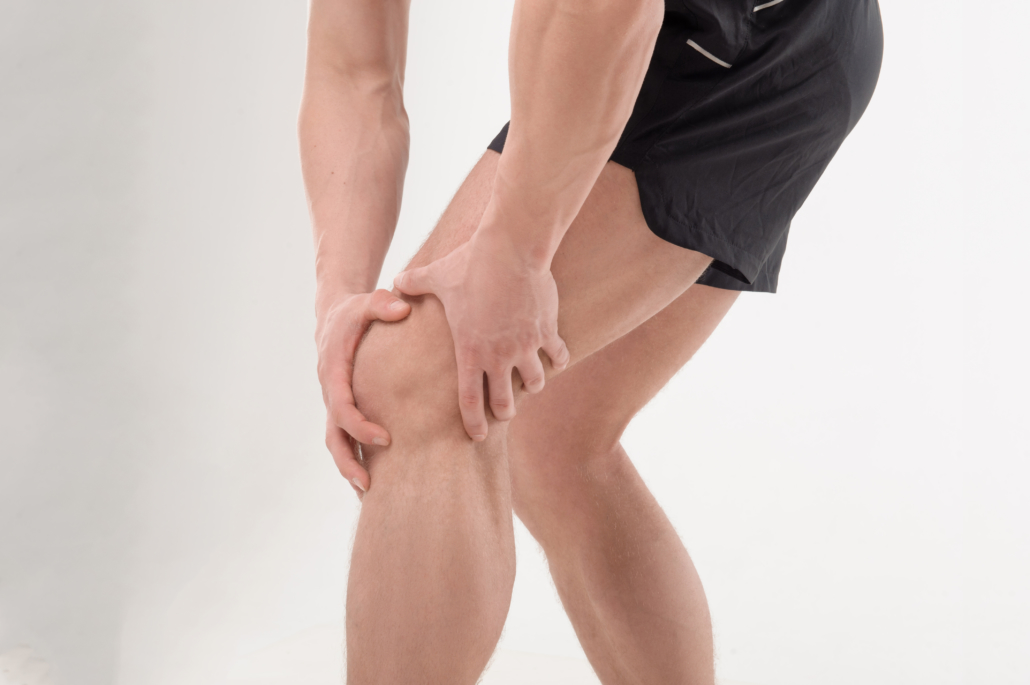
What is a Meniscus Tear?
September 2, 2022 in Article /by Alta HealthMeniscus tear causes, symptoms and treatments
The meniscus is a section of shock-absorbing cartilage that is located between the thigh bone (femur) and the shin bone (tibia). It is split up into two parts: the medial meniscus and the lateral meniscus. Its role is to act as a shock absorber, supply nutrition to the knee, distribute load safely and allow for movement at the knee joint.

How does a meniscus tear happen?
A meniscus tear usually occurs as a result of a rapid twisting of the knee during a weightbearing movement. There are two major types of meniscus tears: acute and degenerative. Acute meniscus tears usually occur in a single leg weightbearing position, such as when changing direction or pivoting. Therefore, meniscus tears are common in change of direction sports such as basketball, football, rugby and tennis.
Degenerative meniscus tears usually occur in elderly populations, as a result of wear and tear of the knee joint. These tears can occur without the twisting mechanism of an acute meniscus tear.
The severity of a meniscus tear is graded on a scale of one to three. A Grade 1 tear is a small tear that does not extend to the articular surface – or the top – of the meniscus. Grade 2 is a larger tear in the meniscus that also does not extend to the articular surface. A Grade 3 tear will intersect the articular surface.
The shape of the tear is also important when classifying the tear. There are six distinct types of meniscus tear shapes. The larger tears and tears of abnormal shape are higher grade and therefore more severe. These tears are also more likely to require surgical intervention to be repaired.
Meniscus tear symptoms
The symptoms of a meniscus tear are dependent on the graded severity and the shape of the tear. The symptoms usually include pain and swelling, knee instability, a clicking or grinding noise when the injury occurs, knee locking or catching, and pain when standing on the affected limb.
Diagnosing a meniscus tear
A meniscus tear is usually diagnosed by a Physiotherapist or Orthopaedic Specialist. There are two meniscus tear diagnostic assessments that may be used: the McMurray’s Test and the Apley’s Grind Test. A positive test is indicated if pain is present, and the knee is catching or grinding during the test. If this assessment is considered positive, an Magnetic Resonance Imaging (MRI) referral is prescribed to examine the severity of the tear. If the assessment is considered negative, further investigation may be needed.
How are meniscus tears treated?
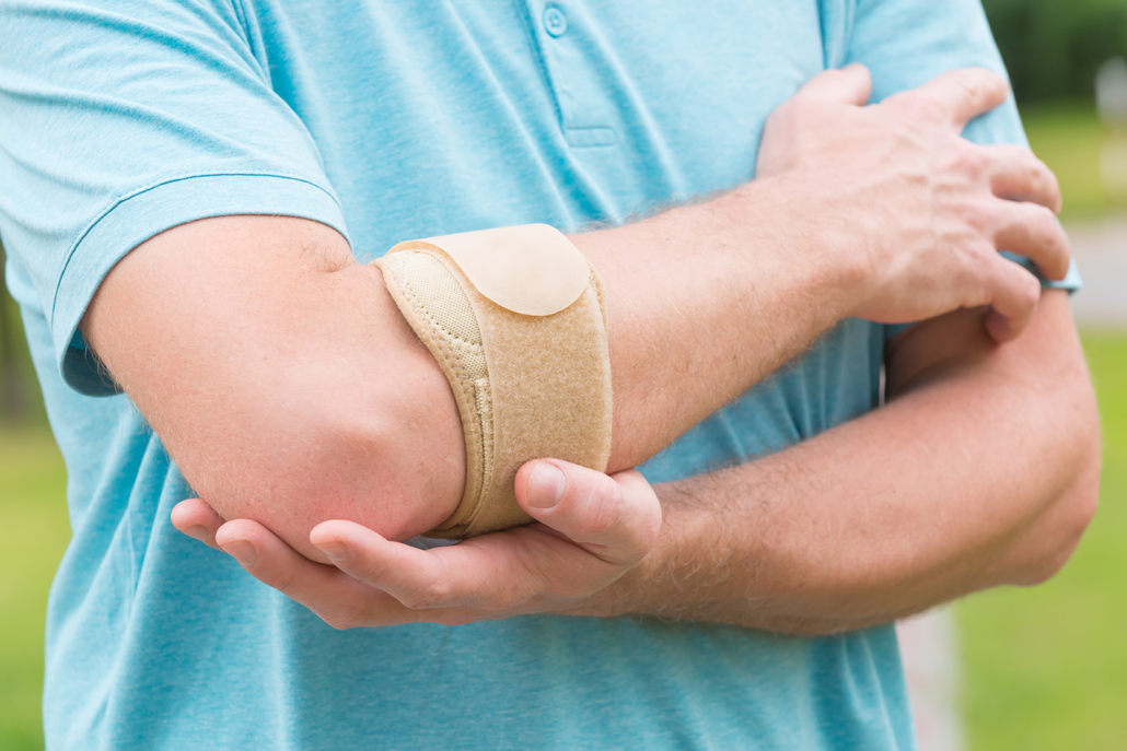
Meniscus tears are treated depending on their severity. The initial treatment of a meniscus tear involves Resting, Icing, Compressing and Elevating (RICE), as well as bracing or taping to stabilise the knee and taking anti-inflammatory medication. Consult a Physiotherapist to improve the knee’s range of motion and reduce pain and inflammation.
How to strengthen the meniscus after a tear
Once pain and swelling has reduced, it is best to consult an Exercise Physiologist (EP). An EP will assess the injury and prescribe an individualised exercise rehabilitation program to re-stabilise the knee joint and improve strength in the muscles surrounding the knee. An EP will also provide an eventual return to sport and work rehabilitation plan.
Some meniscus tears will heal on their own due to a low severity of injury and the favourable location of the meniscus for blood flow. More severe and abnormally shaped tears may require surgical intervention to be repaired appropriately.
How can VALD help?
VALD’s suite of human measurement technologies includes a range of systems which can assist in diagnosing and rehabilitating meniscus tears.
The ForceDecks Dual Force Plate System allows practitioners to gather real-time feedback on relevant metrics, such as single leg and double leg jump height, squat mechanics, and balance and stability assessments.
The NordBord Hamstring Testing System can be used to measure hamstring strength and imbalance in various positions, providing an understanding as to how the muscles surrounding the meniscus injury are progressing.
The ForceFrame Strength Testing System is a fully adjustable system which is used to test isometric strength in upper and lower body positions, including the knee and hip muscles, and joints which are vital to a meniscus tear recovery.
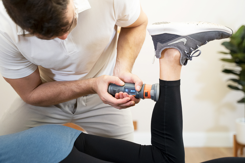
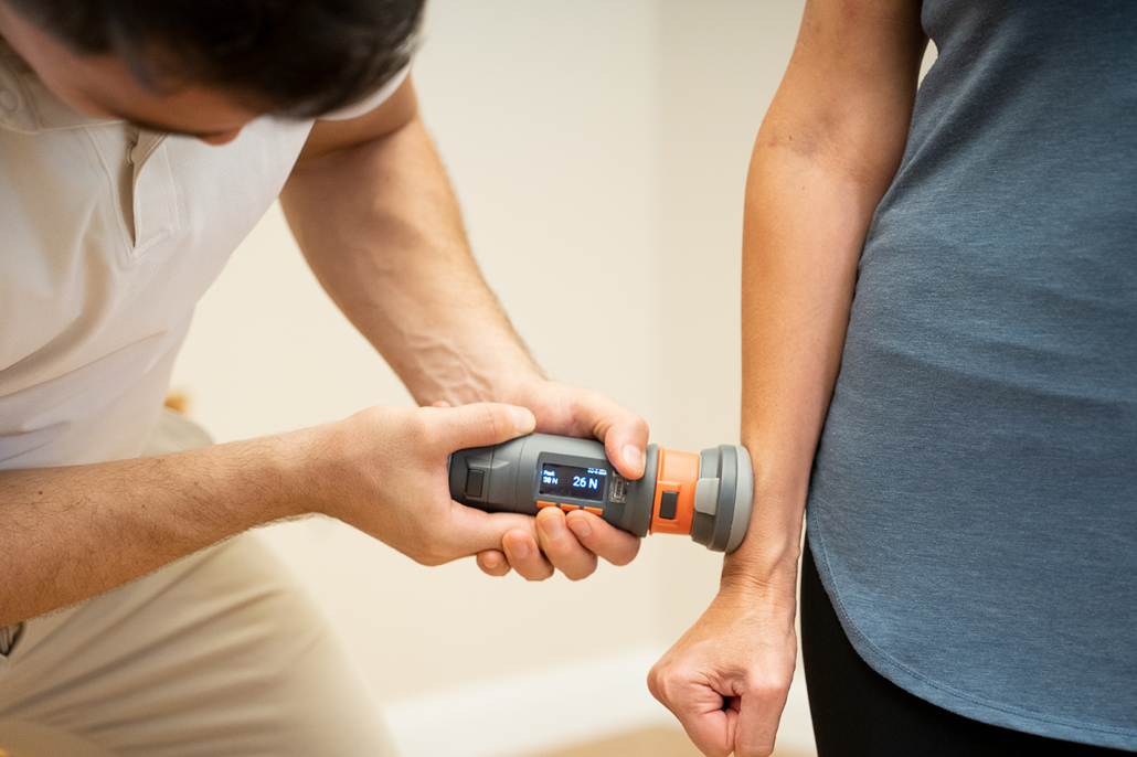
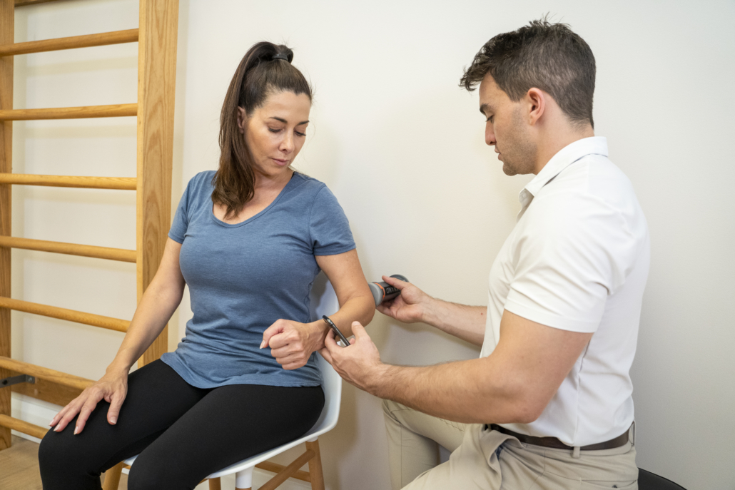
VALD also recently launched a handheld dynamometer and inclinometer named DynaMo capable of performing more than 300 strength and range of motion tests. These tests can be utilised to assess hamstring and quadriceps strength, both of which are important metrics to monitor throughout the rehabilitation process.
To learn more about DynaMo and how dynamometers can be used in the treatment of conditions like a meniscus tear, visit: valdhealth.com/dynamo.